Treatment of Blocked Fallopian via IVF
Introduction
Fallopian tubes are female reproductive organs which connects the ovaries to the uterus. Each month during ovulation, the fallopian tubes carry the egg from an ovary to the uterus. If a fallopian tube is blocked, the passage for sperm to get to the eggs, as well as the path back to the uterus for the fertilized egg is blocked.Symptoms
Common reasons for blocked fallopian tubes include scar tissue, infection, and pelvic adhesions. Blocked fallopian tubes can lead to mild, regular pain in the abdomen.Effect on fertility
Blocked fallopian tubes are a common cause of infertility. If both tubes are fully blocked, pregnancy without treatment will be impossible. If the fallopian tubes are partially blocked, you can potentially get pregnant. However, the risk of ectopic pregnancy increases.Diagnosis
There are several medical tests that can detect some type of blockage.- Hysterosalpingogram (HSG)
- Sonohysterography
- Chromotubation
Treatment
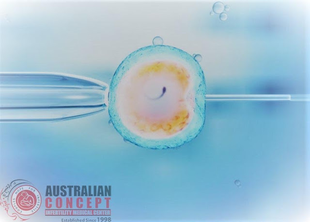
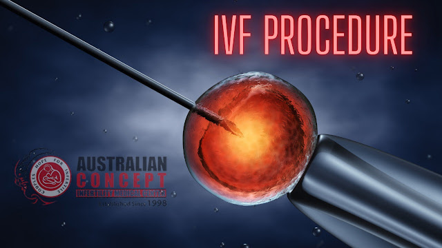
Blocked fallopian tubes prevent natural conception, What is in vitro fertilization (IVF) can bypass the tubes. During IVF, the ovaries are stimulated to produce several eggs, which are then retrieved using a short procedure under anaesthesia. The eggs are then fertilized in the laboratory, and fertilized embryos are placed into the uterus through the cervix where they can implant and grow, so the fallopian tubes are bypassed altogether.The reasons for this result mainly include the following. Water in the hydrosalpinx can return to the uterine cavity and wash the embryo away. Meanwhile, the water contains endotoxins, which is toxic to the embryos. The level of integrin αvβ3 decreases, causing the endometriosis to decrease. This would affect embryo implantation. These patients had a lower embryo implantation rate, clinical pregnancy rate, and higher abortion rate. Therefore, it is very important to treat hydrosalpinx before IVF Treatment.
Therefore, a blocked fallopian tube and no fluid flow to the uterine cavity to provide a relatively good uterine environment for embryo transfer is a problem that needs to be solved before embryo transfer. In order to avoid multiple laparotomy or laparoscopic surgery, and reduce the risk of anaesthesia and surgery, modern fallopian tube obstruction technology has emerged as the times require.
Rosenfield reported the first case of tubal embolization through the hysteroscopic placement of a micro insert, which was an Essure (Conceptus). The patient had a 6.5-year history of infertility. A hysterosalpingogram revealed a left hydrosalpinx. The patient was offered surgical intervention to treat the hydrosalpinx to potentially improve the chance for successful pregnancy through IVF-ET. However, the 31-year-old nulligravid woman underwent laparoscopic left oophorectomy for a persistent 6-cm complex adnexal mass, because of a body mass index of 50.5 kg/m2. In order to avoid the previously recounted risks of general anaesthesia and laparoscopy in this morbidly obese woman, the patient elected to undergo hysteroscopic placement of a micro insert. After the in vitro fertilization, the patient had a good outcome. Hence, it is a reasonable method to find the opening of the fallopian tube by hysteroscopy and place a micro insert into the tubal cavity to block the fallopian tube. Compared with x-ray mediated tubal embolization, the advantage of this procedure was that it is similar to the fallopian tube intubation fluid. It can be completed under paracervical nerve block anaesthesia in an outpatient clinic, without contact with radiation therapy. Furthermore, the operation has no effect on ovarian function. Therefore, it is an ideal method for patients who need tubal embolus due to the test tube. However, hysteroscopic tubal embolization is not yet a mature procedure, and loss during the placement of micro coils is the most difficult event to explain. If the micro-coil falls into the uterine cavity, it will be easy to remove. However, it would be difficult to remove if it enters the ventral ampulla of the fallopian tube or abdominal cavity. Therefore, we need to explore the correct placement method to eliminate such events.
Methods
Interventional embolization: Interventional embolization could be completed by the outpatient procedure. After routine examination of chlamydia, mycoplasma and leucorrhea, patients with normal parameters received interventional embolization. Placed in the lithotomy position, patients lay on the digital subtraction angiography with routine disinfection, draping and chamber testing. Then fluoroscopically-guided 7 F oviduct catheter was inserted into cornua uteri of hydrosalpinx across the cervical canal. Under the guidance of 0.018 in the guidewire, 3 F microcatheter was gently inserted into interstitial section and isthmus of the fallopian tube, after which guidewire was slowly pulled out. According to the length of inserted microcatheter and the thickness of the fallopian tube, the proper micro coil was selected and released via 3F catheter in the drive of a guidewire (trying to locate the near-end of a micro coil in the cornua uteri next to interstitial section). Contralateral fallopian tube of patients with bilateral hydrosalpinx was treated with the same method. Finally, Hystero-Salpingography (HSG) was conducted to verify the effect of embolization. After a month, HSG was performed again to verify the embolization and patients without therapeutic effect need another embolization.Therapeutic evaluation of embolism:
- Significant effect:
Microcoil located in fallopian tube with a length of near-end to oviduct aperture<10 mm;
- Effective:
The length of near-end to oviduct aperture was 10-50 mm;
- Invalid:
Contrast
agent permeated to the far-end cross micro-coils; micro coil shifted to the
far-end or fallen off to uterine cavity.


Comments
Post a Comment